Equipment:
- Microscope with stage FP-90 (Mettler-Toledo) for thermo-optical analysis with DSC
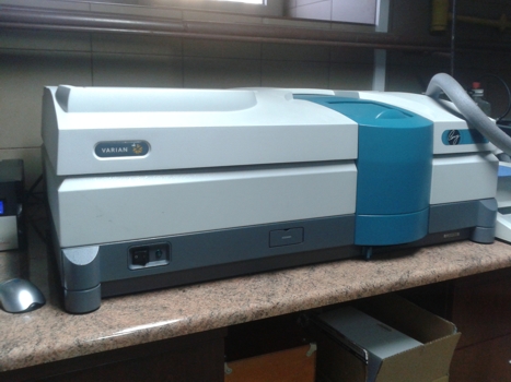
- UV-VIS-NIR spectrometer (Varian Inc. Carry 5000) (175 – 3300 nm range) with integrating sphere
Parameters:
Apparatus wavelength range 175 - 3300 nm. Its equipment includes:
- integrating sphere for transmittance and reflectance measurements of highly disperse samples,
- polarizer/depolarizer,
- attachment for reflectance measurements under different angles,
- Peltier attachment for liquid samples measurements in temperature range 0-100°C,
- set of holders and diaphragms of various shapes and sizes for solids measurements (plates, foils, films),
- holders for cells with various optical path (0.1 - 100 mm),
- mass transporter for analysis of heterogeneous materials transmittance (spatial resolution 1 mm)
- Photoluminescence Spectrometer FLS980 (Edinburgh Instruments) with inverted optical microscope (TE2000-U)
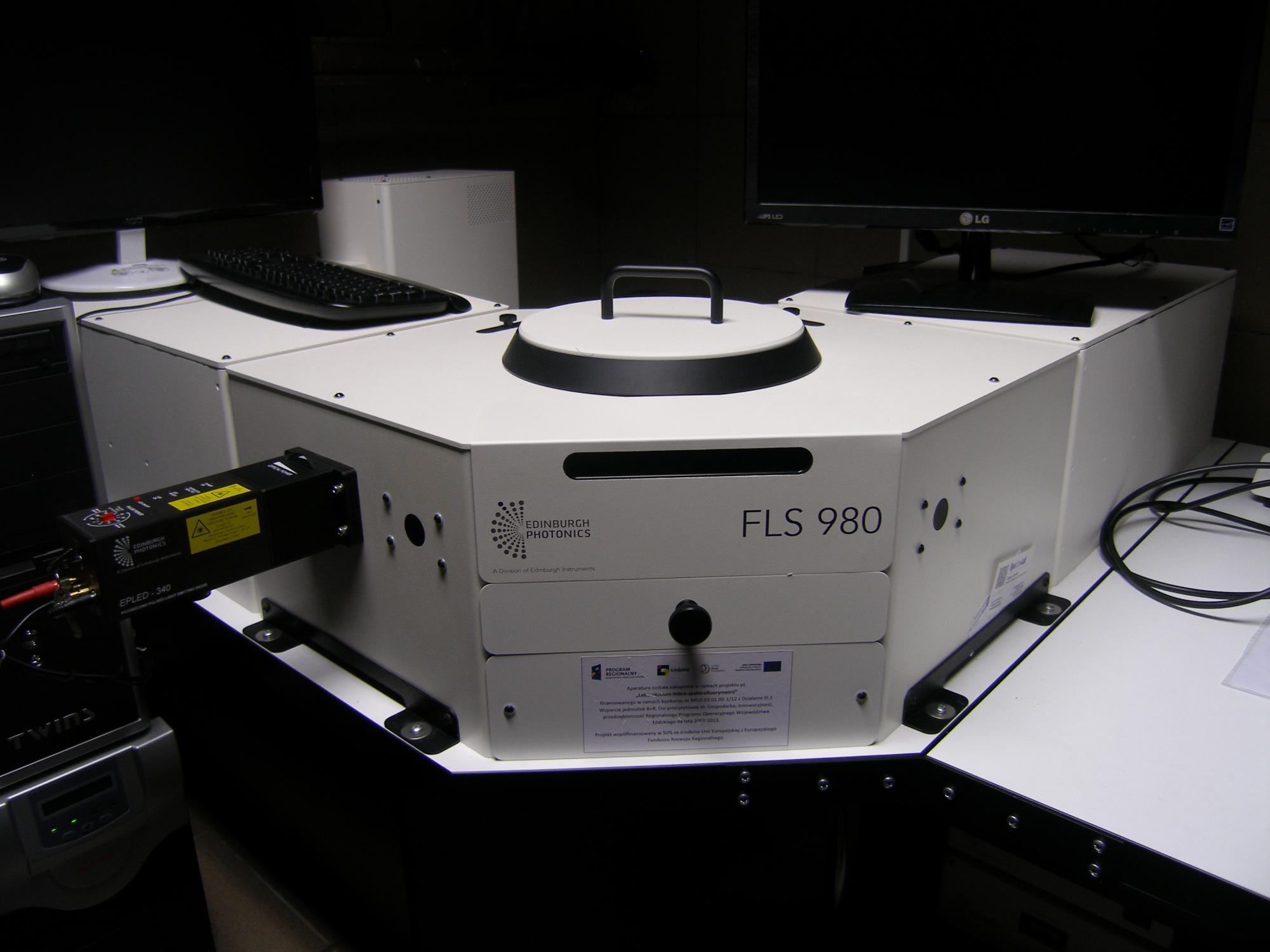

Apparatus was purchased in frame of the project „Laboratory of micro-spectrofluorymetry” applied in RPLD.03.01.00-1/12 contest. Project is founded in 50% by European Union from European Regional Development Found in frame of National Cohesion Strategy. Duration time: 01.11.2012 - 31.08.2013 r.
Parameters:
Excitation source: 450W, Ozone free Xenon Arc Lamp, detector: photomultiplier R928P, spectral range 200-870 nm, double grating excitation and emission monochromators, Czerny-Turner type. The FLS980 is modular spectrofluorimeter for measuring steady state and time resolved luminescence spectra of liquid and solid samples. The NIKON ECLIPSE TE2000-U inverted microscope is connected to the spektrofluorimeter. Additional equipment includes:
- front face sample holder with external adjustment for accurate sample positioning. The accessory comes with inserts for cuvette, film/slide clamp and holder for micro-samples,
- the integrating sphere allows absolute quantum yield, reflectance and chromaticity measurements to be taken. It comes with sample holders for liquids, powders and films,
- motorized high quality Glan-Thompson polarizers,
- excitation source for time correlated single photon counting operation mode (TCSPC) is pulsed light emitting diode: emission 340nm, pulse width below 750 ps, repetition rate 10MHz.
Contact person: Marcin Kozanecki, Gabriela Wiosna-Salyga, Ewelina Witkowska
FLS980 connected to optical microscope is ideal system for demanding applications in the broad areas of photophysics, photochemistry, biophysics and material science. Spectrofluorimeter is able to measure steady state luminescence and excitation luminescence spectra in the ultraviolet to near infrared spectral range in right angle and front-face geometry. The front-face mode is especially useful for highly concentrated, opaque, or solid samples such as blood, paint, optical brighteners, living cells, polymer films or phosphors. The steady state fluorescence and excitation fluorescence anisotropy may be obtained from polarized excitation and emission spectra. The analysis of such measurement is powerful tool for directly studying the molecular rotation of the fluorophore and is often employed to protein folding and membrane fluidity studies. The absolute method for fluorescence quantum yield measurements is becoming more widely used than the relative method, as it does not require a quantum yield standard, is readily applicable to liquids, films and powders. The chromaticity option of the FL980 calculates and displays color co-ordinates for CIE1931 and CIE1976, and also produces luminance values.
Time resolved emission spectroscopy and global analysis allow to understand complex excited state dynamics and define the processes which take place after excitation of molecule (e.g. proton/electron transfer, solvent relaxation, excimer formation). An unquestionable advantage of our system is connection between FL980 and inverted microscope. Fluorescence microscopy has long been established as an essential tool in biology and the biomedical sciences, as well as in materials science. It detects the fluorescence signal from the molecules which are excited by specific wavelengths of irradiating light and generates images of illuminated specimen. When the specimen doesn’t exhibit autofluorescence the fluorochromes are added. Various fluorescent indicators are available to study many physiologically important chemicals such as DNA, calcium, magnesium, sodium, pH and enzymes. Fluorescence-microscopy technique is useful for seeing structures of host-guest systems and studying the mechanism of energy transfer.