Equipment:
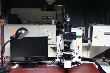
- Raman Spectrometer (Jobin-Yvon T64000) with Olympus Confocal Microscope
Parameters:
Dispersive spectrometer T-64000 (Jobin-Yvon) is a very “flexible” platform for Raman analysis. Two fundamental work modes may be used: triple – three-grating monochromator (1800 lines/mm) offers high spectral resolution (up to 0,5 cm-1) and very well rejected laser signal giving an access to low frequency range down to 10 cm-1; single – only one grating monochromator is used (optionally 600 or 1800 lines/mm); this option is useful for fast analysis in broad range of frequency. The classical Raman experiments in so called T-configuration (an exciting and a scattered beams are perpendicular one to each other) are realized in large macro chamber with removable walls. This makes possible measurements of large samples and usage of different accessories. Currently Department of Molecular Physics offers the Peltier heating-cooling system (273-373 K) dedicated to liquid and gel-like samples. The micro-Raman measurements are realized in back-scattered configuration and both transparent as well as opaque samples may be analyzed. T-64000 spectrometer is coupled with real confocal microscope (Olympus BX-40) equipped with motorized XY stage. Such solution allows for mapping with lateral spatial resolution below 1 um and the depth profiling with resolution closed to 2.5 um. An unique Helq/N2lq home made cryostat dedicated to described system is equipped with internal electrical connections giving a possibility of simultaneous measurements of Raman spectra and electrical conductivity. The temperature range accessible with usage of this cryostat starts from c.a. 4 K. The high temperature measurements (up to 573 K) may be realized with heating-stage (SEMIC Bioelektronika). Both types of experiments classical as well as micro-Raman may be realized with different excitation wavelengths: 457.9, 476.5, 488.0, 496.5, 514.5, 647.1, 676.4 nm. The polarizing measurements are also accessible for both experiments.
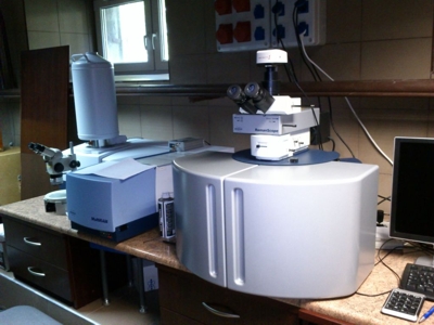
- FT-Raman Spectrometer (Bruker RamanScope III) with Olympus Microscope
Parameters:
Fourier transform Raman spectrometer MultiRAM (Bruker GmbH) is a powerful analytical tool provides a spectral range of 3600 – 100 cm-1 (Stokes shift). The standard light source is a diode-pumped, air-cooled Nd:YAG laser (1064 nm; 1000mW). This spectrometer is combined with microscope RamanScope. Such solution offers a spectral resolution equal 1 cm-1 and a lateral spatial resolution below 10 um. Motorized XYZ stage gives possibility of mapping and depth profiling.
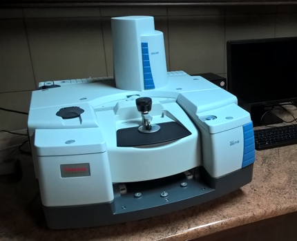
- FT-IR spectrometer (Thermo Scientific Nicolet is50) (12 000 – 50 cm-1 range)
Parameters:
A Thermo Scientific Nicolet is50 Fourier Transform Infrared Spectrometer FTIR allows to collect data over a wide spectral range (12 000 – 50 cm-1). It is possible to obtain infrared spectra with a resolution of 0.125 cm-1. Spectrometer is equipped with two sources. Available detectors are: InGaAs photodiode (indium gallium arsenide) - measurements in the near-IR range, DTGS pyroelectric (deuterated triglycine sulfate) and the MCT-B photoconductive (mercuric cadmium telluride) cooled liquid nitrogen - to record spectra in the mid-IR, DTGS-PE (deuterated triglycine sulfate with polyethylene window) to detect spectra in the far-IR range. The measuring system is equipped with an Automatic Beamsplitter Exchanger (ABX) which provides multi-range spectrometry without having to manually swap optics. When you choose required spectral range, the is50 ABX loads the correct beamsplitter into the interferometer. Spectrometer Thermo Scientific Nicolet IS50 can work in two configurations: transmission mode - using the transmission method we can analyze samples in all states of matter (liquids, solutions, solids and gases), after preparing the material. The method is standard, simple and inexpensive. Measurements of corrosive and caustic samples are possible. The transmission mode is limited by thickness of samples (max. 10-12 microns), this method provides information about the whole sample; ATR method (attenuated total reflectance) with a diamond crystal - information is obtained from the sample surface. We can measured materials characterized by strong absorption, providing analysis of samples in their natural states. In the ATR module there is no thickness limitation.
Contact person: Marcin Kozanecki, Paulina Filipczak
Vibration spectroscopy is one of the most popular analytical techniques for both qualitative and quantitative analysis. The vibrational spectrum of a substance is like a fingerprint - characteristic of a given substance and unique. This allows for unambiguous determination of the composition of the analyzed sample. Using the classic methods of the standard curve or internal standard, it is also possible to perform quantitative analysis of complex systems. The Department of Molecular Physics specializes in the use of vibrational spectroscopy methods in material research. The interests of the Department's employees include both organic, inorganic and hybrid materials. The most frequently conducted research includes the characterization of intermolecular interactions and phase analysis (including phase transitions), although the employees of the Department are also experienced in the study of chemical reactions. Currently, the term vibration spectroscopy covers many research techniques that differ significantly in the measurement methodology, the equipment used and the scope of the studied phenomena.
micro-Raman spectroscopy
Modern hi-tech materials requires advanced analytical methods to be well described. The micro-Raman spectroscopy is an unique technique offers a possibility of simultaneous analysis of both spectral and structural properties of measured sample. One of the most important advantage of micro-Raman spectroscopy is also possibility of fast analysis of the samples without any special pre-treatment, independently on their shape, form and state. Due to that Raman spectroscopy is a very useful analytical techniques in many scientific as well as industrial fields of activity such as chemistry, pharmacy, food science, physics, material science and technology, nanotechnology, geology and others. Recently this technique has been also successfully applied in medicine as diagnostic method. The main application of micro-Raman spectroscopy may be grouped into two main domains:
1. Chemical analysis:
- Quantitative and qualitative analysis
- Analysis of molecular conformations and configurations
- Analysis of intermolecular interactions
- Control of chemical reactions
2. Material science analysis:
- Investigations of molecular order and orientation in materials
- Investigations of phase transitions and other physical processes
- Investigation of heterogeneous materials
- Analysis of internal stresses and their distribution
Applications of FTIR:
- Identification of inorganic compounds and organic compounds
- Identification of components of an unknown mixture
- Analysis of solids, liquids and gasses
- Determination of the purity of compounds
- Control of chemical reactions
- Analysis of the surface composition
- Quantitative analysis
- The study of intermolecular interactions
Selected publications:
- “Spontaneous versus Stimulated Surface-Enhanced Raman Scattering of Liquid Water” – P. Filipczak, M. Pastorczak, T. Kardaś, M. Nejbauer, Cz. Radzewicz, M. Kozanecki – J. Phys. Chem. C 2020 doi: 10.1021/acs.jpcc.0c06937
- “Water structure and hydration of polymer network in PMEO2MA hydrogels” – K. Piechocki, M. Kozanecki, J. Saramak – Polymer 210 (2020) 122974; DOI: 10.1016/j.polymer.2020.122974
- “Molecular Spectroscopy—Experiment and Theory. From Molecules to Functional Materials.” Chapter 8: “Vibrational spectroscopy in analysis of stimuli-responsive polymer-water systems.” – M. Kozanecki, M. Pastorczak, K. Halagan – Eds. A. Kolezynski, M. Krol; Springer Nature Switzerland AG 2019 – book series: Challenges and Advances in Computational Chemistry and Physics” vol. 26. pp. 223-271.
- „Surface-enhanced Raman spectroscopy (SERS) in cotton fabrics analysis.” – D. Puchowicz, P. Giesz, M. Kozanecki, M. Cieślak – Talanta 195, 516–524 (2019)
- „The influence of cellulose derivatives on water structure in gypsum.” – A. Czaderna, A. Kocemba, M. Kozanecki, M. Mucha, P. Mróz – Construction and Building Materials 160, 628–638 (2018)
- „Raman spectroscopy study on influence of network architecture on hydration of poly(2-(2-methoxyethoxy)ethyl methacrylate) hydrogels.” – M.N. Olejniczak, M. Kozanecki, J. Saramak, M. Matusiak, S. Kadlubowski, K. Matyjaszewski – J. Raman Spectr. 48, 465-473 (2017)